Ultrasound machines and software
Because every image counts
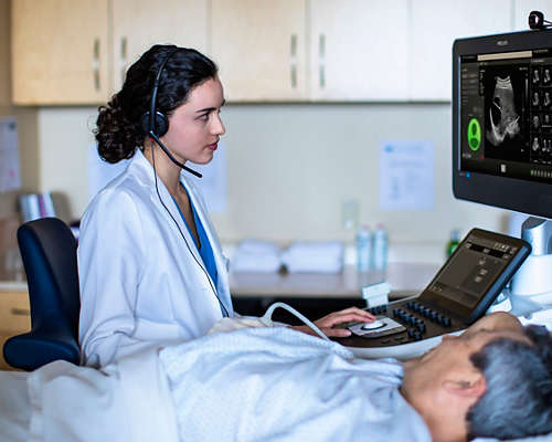
Philips ultrasound solutions connect technology, clinicians and patients to empower patient-focused care and elevate the healthcare experience. Our comprehensive portfolio – ranging from ultra-mobile handheld devices to premium cart-based systems – is designed to empower clinical confidence, with innovations that enhance imaging accuracy and performance. We’re committed to designing sustainable solutions for reliability, scalability and ease-of-use. Paired with shared architecture and comprehensive service programs, our solutions deliver lifetime value to you and your patients.


Shared system DNA enhances walk-up usability
Philips ultrasound machines feature a common user experience across the platform, so you can choose the best combination for your clinical needs. This shared DNA, combined with thoughtful features that promote walk-up usability, streamlines training, supports staffing flexibility and simplifies reporting workflow. In addition, many of our transducers can be used on a variety of systems, allowing for greater flexibility in managing your transducer inventory.
Featured products in Ultrasound machines and software
-
5500CV
Philips Compact Ultrasound System 5500CV brings full functionality and first-scan answers to you, wherever you are. Offering a feature-rich core, a range of diagnostic quality solutions, enhanced cleanability and wireless connectivity and reporting, Philips Compact Ultrasound System 5500CV is one of the most reliable and robust compact systems on the market. The Compact 5500CV system won an iF Design Award and a Red Dot Product Design award in 2021 for its easy mobility.
795141 -
5300 series
Philips Compact Ultrasound System 5300 series brings full functionality and supports quick, confident decision making, wherever you are. Designed for the many different clinical environments in general imaging, point-of-care, and obstetrics and gynecology, the Philips Ultrasound 5300 model offers a feature-rich core and a versatile range of diagnostic solutions - all built into a highly mobile, cleanable, easy-to-use system. The Compact 5300 series won an iF Design Award and Red Dot Product Design Award in 2021 for its easy mobility.
795136 -
EPIQ Elite
Philips EPIQ Elite ultrasound features an exceptional level of clinical performance, workflow, and advanced intelligence to meet the challenges of today’s most demanding practices. The EPIQ Elite platform brings ultimate solutions to ultrasound, with clinically tailored tools designed to elevate diagnostic confidence to new levels.
795098 -
EPIQ CVx
The EPIQ CVx is a dedicated cardiac ultrasound solution which brings significant advancements in functionality. This helps you deliver better care through higher processing power, exceptional imaging with more clarity & sharpness, improved exam efficiencies, complemented by the proven, robust quantification capabilities of TOMTEC.
795231 -
Affiniti 70
Philips understand the challenges you face. From a changing patient population to the need to maximize resources, the challenges can be considerable. Philips Affiniti helps you overcome these daily challenges so you can provide the best possible care for your patients every day. Affiniti offers a powerful combination of performance and workflow advances to enhance diagnostic confidence making it the choice of clinicians worldwide.
795210 -
Affiniti 50
Choosing a new ultrasound system is all about balance. You need accurate diagnostic information quickly, a simplified yet intuitive user interface, and easy access to critical features, along with an ergonomic design and the latest technology.
795208


- 0
Extend your team without expanding it
Philips Collaboration Live* enables secure talk, text, screen share and video stream functions, allowing users to remotely access expertise directly from the ultrasound system. The Collaboration Live solution can be used to provide real-time remote clinical diagnosis,** decision support and training on care protocols. *Collaboration Live requires VM 7.0.5 or higher. Remote diagnostic use requires VM 9.0 or higher.
**Accessible via compatible clients including iOS, Android, Chrome web browser and Windows with release 9.0 or higher.
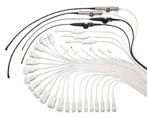
High-resolutions transducers for swift and precise imaging
Philips offers a wide range of transducers for excellence in echocardiography, doppler and sonography. They feature intelligent imaging technology for 2D, 3D, 4D, flat, static and moving imaging along with an ergonomic design for comfortable scanning. For example, leading-edge xMATRIX technology enables quick and easy volume acquisition, supports multiple interrogation capabilities, and provides views not possible with 2D imaging.
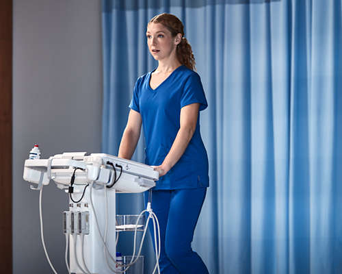
It’s compact without compromise
Compact Ultrasound System 5000 series brings full functionality and supports quick, confident answers to you, wherever you are. The Compact 5000 Series offers a feature-rich core and a versatile range of diagnostic solutions – all built into a highly portable, cleanable, easy-to-use system.

Ultrasound capabilities in the palm of your hand
Lumify handheld ultrasound helps you diagnose and treat patients wherever they are – from the ambulance to the operating room and everywhere in between. With its rugged, military-grade hardware and innovative software, you can acquire critical clinical data quickly and easily. Lumify incorporates Reacts technology so that you can instantly collaborate with colleagues anytime and anywhere.
Technologies and innovations
-
xMATRIX transducer technology enables scanning in two planes simultaneously. Available in 13 scanning modes, it enhances image clarity and eases workflow to make exams faster and easier for both clinicians and patients.
-
Available on Philips ultrasound machines, Flow Viewer defines vasculature with a 3D-like appearance and reduced flash artifact.
-
Available on Philips ultrasound transducers, MicroFlow Imaging (MFI) and MicroFlow HD (MFI-HD) detect slow and weak blood flow anatomy in tissue.
-
![Technology Maximizer]()
Learn how Technology Maximizer secures all your Philips imaging equipment with the same technology release level**, reducing maintenance complexity and simplifying lifecycle management across hospital departments.
-
Philips AI-based ultrasound solutions integrate into everyday clinical workflows and are designed to make it easier and faster for clinicians to acquire, select, measure and report accurate results.


